Pulmonary Valve Atresia
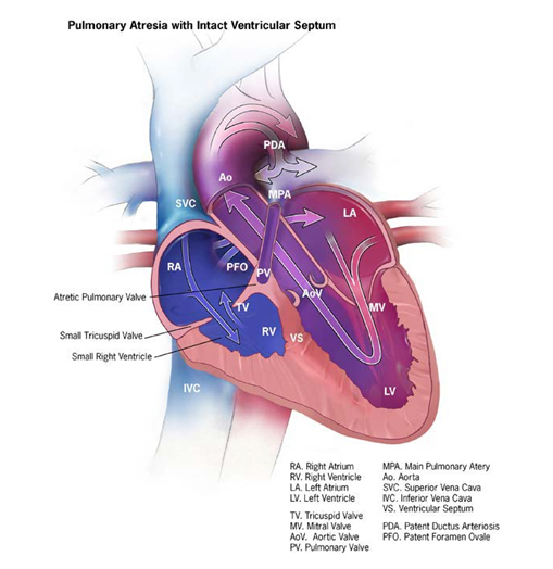
Pulmonary valve atresia is a structural heart anomaly characterized clinically by cyanosis and anatomically by an imperforate pulmonary valve that blocks completely the flow of blood through the right ventricular outflow tract. The atresia can take the form of a membrane – because the valve failed to form – or of an imperforate muscle structure (see Fig. 17).
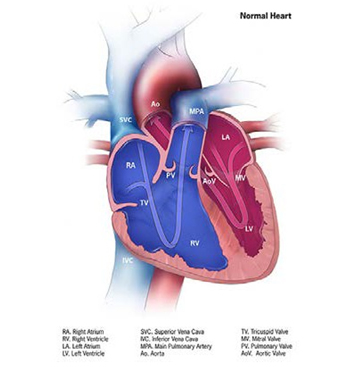
Fig. 17. Pulmonary valve atresia


- An imperforate pulmonary valve.
- A ventricular septum that is intact or can present a ventricular septal defect: This is a very important detail to look for and describe – the panel above in Fig. 17 shows the form of pulmonary atresia with an intact ventricular septum.
- An underdeveloped right ventricle and possibly a small or narrowed tricuspid valve, especially if the ventricular septum is intact.
Diagnosis
Prenatal. Pulmonary valve atresia can be suspected prenatally based on ultrasonography but must be confirmed postnatally.
Postnatal. Infants with pulmonary atresia (with or without ventricular septal defect) typically present early in the neonatal period with low oxygen saturation and cyanosis, which worsens over time as the ductus closes. Some infants might also have massive cardiomegaly. Because pulmonary atresia causes low blood oxygen saturation, newborn screening with pulse oximetry can help with early detection of these cases. Echocardiography is the key diagnostic procedure, although other imaging techniques, including catheterization, might be necessary to fully guide management and care.
Clinical and epidemiologic notes
Pulmonary atresia with intact ventricular septum is often isolated but can be associated with unrelated anomalies and syndromes as well as with other intracardiac anomalies, especially those that involve the right side of the heart.
Pulmonary atresia with ventricular septal defect can be associated with deletion 22q11, unlike the form with intact ventricular septum. Because of this association, look for other birth defects, including cleft palate and internal anomalies.
Checklist for high-quality reporting
| Pulmonary Atresia – Documentation Checklist |
Describe in detail the clinical and echocardiographic findings:
Look for and document extracardiac birth defects: In deletion 22q11, the heart anomaly can be associated with several internal and external anomalies, including cleft palate, spina bifida, vertebral anomalies, or other defects. Report whether specialty consultation(s) were done,such as whether the diagnosis was made by a paediatric cardiologist, and whether the patient was seen by a geneticist Report any genetic testing and results s (e.g. chromosomal studies, genomic microarray). |