Transposition of Great Arteries
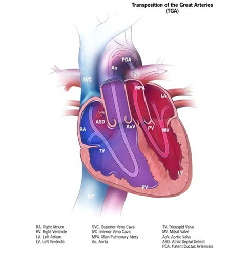
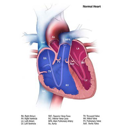
d(dextro)-transposition of the great arteries (d-TGA) is a structural heart anomaly characterized clinically by cyanosis (usually) and anatomically by an abnormal origin of the great arteries, such that the aorta exits from the right ventricle (instead of the left) and the pulmonary artery exits from the left ventricle (instead of the right) (see Fig. 15).
Fig. 15. Transposition of great arteries


- Aorta exits from the right ventricle (instead of the left) and the pulmonary artery exits from the left ventricle (instead of the right).
- Ventricular septal defect might or might not be present, and if present, should be documented and reported.
- Terms to look for and to document (to help the coder and central reviewer distinguish d-TGA from other forms of transposition) include double outlet right ventricle, levo (l)-transposition of the great arteries, heterotaxy and single ventricle.
- Echocardiography is the evaluation that in most cases provides all the information required for a precise diagnosis.
Diagnosis
Prenatal. d-TGA can be suspected prenatally, but prenatally diagnosed or suspected cases should be confirmed postnatally.
Postnatal. Infants with d-TGA present in a variety of ways, most commonly with cyanosis that worsens as the ductus closes, or occasionally also with heart failure (usually when a large ventricular septal defect is present).
Newborn screening via pulse oximetry, which is based on the non-invasive detection of low blood oxygen saturation, can detect many cases of d-TGA even before overt clinical symptoms.
Clinical and epidemiologic notes
As noted, infants present typically early after birth with cyanosis, which does not improve much or at all by providing oxygen (the problem is not in the lungs but in the abnormal circulation of blood due to the transposed arteries). As noted, rapid clinical deterioration is expected as the ductus arteriosus closes. d-TGA is most commonly an isolated heart anomaly, but extracardiac anomalies are found in ~10% of cases.
Checklist for high-quality reporting
| d-TGA – Documentation Checklist |
Describe in detail the clinical and echocardiographic findings:
Look for and document extracardiac birth defects: These are not as common as in other conotruncal defects, but can occur. Report whether specialty consultation(s) were done; for instance, whether the diagnosis was made by a paediatric cardiologist, and whether the patient was seen by a geneticist. Report any genetic testing and results (e.g. chromosomal studies, genomic microarray, etc.). |