SARS-CoV Images
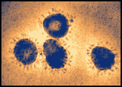
Human coronavirus 229E virus particles, a coronavirus in the same family as SARS-CoV, as seen in a colorized electron microscopic image. Virions contain characteristic club-like projections emanating from the viral membrane.
Image source: F.A. Murphy and S. Whitfield, CDC
Negative stain electron microscopy shows a SARS-CoV particle with club-shaped surface projections surrounding the periphery of the particle, a characteristic feature of coronaviruses.
Image source: C.D. Humphrey, CDC
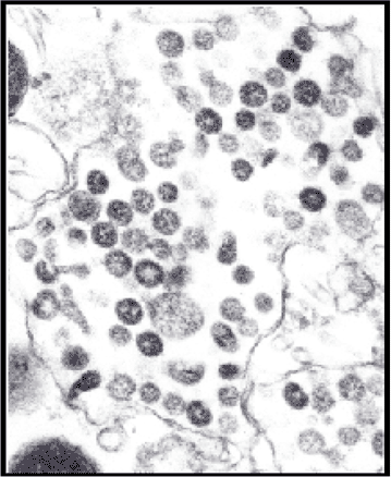
An electron microscopic image of a thin section of SARS-CoV within the cytoplasm of an infected cell, showing the spherical particles and cross-sections through the viral nucleocapsid.
Image source: C.S. Goldsmith, CDC
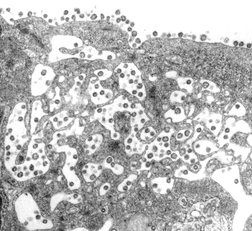
A SARS-CoV-infected cell with virus particles in vesicles, which appear to migrate toward the cell surface and fuse with the plasma membrane, releasing the viral particles. Many of the particles adhere to the plasma membrane, creating a characteristic knob-like appearance on the surface of the cell.
Image source: C.S. Goldsmith, CDC


