4.9g Limb Deficiency: Longitudinal Postaxial (Fibula, Ulna, Fifth Ray) (Q71.30, Q71.5, Q72.30, Q72.6)
‹View Table of Contents
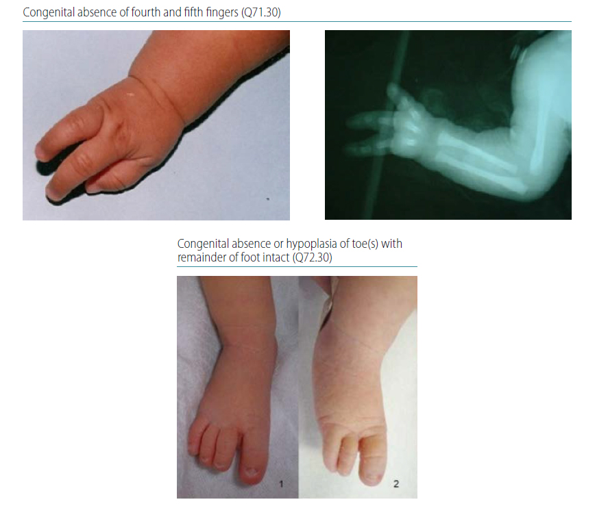
Postaxial limb deficiency is characterized by absence or hypoplasia of the fifth toe/finger (sometimes also including the fourth toe/finger) with or without absence/hypoplasia of the fibula or ulna (see Fig. 4.40). Radiographs are strongly recommended to confirm the findings and characterize the bony anomalies.
Fig. 4.40. Longitudinal postaxial

Relevant ICD-10 codes
Q71.5 Longitudinal reduction defect of ulna
Q72.6 Longitudinal reduction defect of fibula
Absence of fibula
Q71.30 Congenital absence of finger(s) with remainder of hand intact
Q72.30 Congenital absence or hypoplasia of toe(s) with remainder of foot intact
Note: Avoid using the generic Q71, Q72 or Q73 codes for longitudinal postaxial limb deficiencies. These generic codes include otherlimb reduction defects.
When using Q71.30 – congenital absence of finger(s) with remainder of hand intact – or Q72.30 – congenital absence or hypoplasia of toe(s) with remainder of foot intact – be sure to denote which fingers and toes are affected to be able to differentiate from transverse terminal defects.
Diagnosis
Prenatal. Longitudinal postaxial limb deficiencies might be diagnosed or strongly suspected prenatally. However, they can be missed. The distinction from other limb deficiencies is difficult and error-prone. For this reason, a prenatal diagnosis should always be confirmed postnatally.
When this is not possible (e.g. termination of pregnancy or unexamined fetal death), the programme should have criteria in place to determine whether to accept or not accept a case based solely on prenatal data.
Postnatal. The newborn examination can identify a longitudinal postaxial limb deficiency and distinguish it from other limb deficiencies (e.g. longitudinal postaxial defects). An accurate and complete diagnosis requires a detailed physical examination aided by radiography to characterize completely the bony anatomy.
Clinical and epidemiologic notes
Absence or hypoplasia of the ulna typically affects only one arm. With complete absence of the ulna, there is often a marked flexion deformity of the elbow. The hand can be straight or angulated to the ulnar side of the wrist.
Ulnar deficiency is less common than radial deficiency. Ulnar hypoplasia is often associated with radioulnar synostosis (fusion of the radius and ulna), absence of the postaxial digits (fourth and fifth fingers) and fibular deficiency.
Some associations and syndromes described with postaxial defects include the following:
- femur-fibula-ulna complex, characterized by the unilateral absence or hypoplasia of the ulna, femur and fibula;
- ulnar-mammary syndrome (a genetic condition), characterized by deficiencies of the ulna, fibula and postaxial digits; hypogenitalism; and absence of one or both breasts/nipples; and
- Miller syndrome, in which facial differences are associated with postaxial deficiencies of the upper limb.
Distinguishing longitudinal postaxial defects from other limb reduction defects is important because these conditions have different causes and associated anomalies. With careful clinical and radiological examination, the diagnosis of longitudinal postaxial limb deficiencies is possible.
The reported prevalence of postaxial limb deficiencies is approximately 0.45 per 10 000 births.
Inclusions
Q71.30 Congenital absence of finger(s)
Q71.5 Longitudinal reduction defect of ulna
Q72.30 Congenital absence or hypoplasia of toe(s) with remainder of foot intact
Q72.6 Longitudinal reduction defect of fibula
Exclusions
Q71.31 Absence or hypoplasia of thumb
Q71.4 Longitudinal reduction defect of radius
Q71.6 Congenital cleft hand
Q72.31 Absence or hypoplasia of first toe with other digits present
Q72.5 Longitudinal reduction defect of tibia
Q72.7 Split foot
Checklist for high-quality reporting
| Longitudinal Postaxial Defects – Documentation Checklist |
Describe in detail, including:
|
Suggested data quality indicators
| Category | Suggested Practices and Quality indicators |
| Description and documentation | Review sample for documentation of key descriptors:
|
| Coding |
|
| Clinical classification |
|
| Prevalence |
|
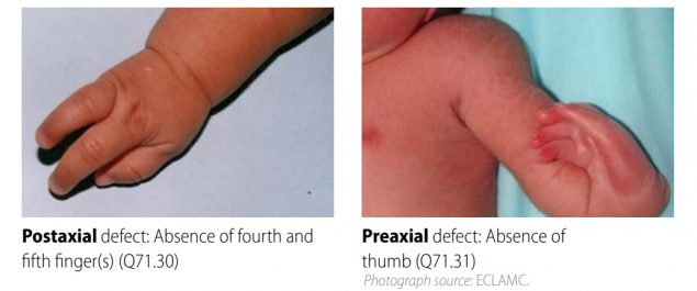
| Key visuals | Distinguishing longitudinal postaxial defects from longitudinal preaxial defects (side-by-side comparison):
 |
Table of Contents
- Chapter 4: Diagnosing and Coding Congenital Anomalies
- 4.1 List of Selected External and Internal Congenital Anomalies to Consider for Monitoring
- 4.2 Congenital Malformations of the Nervous System: Neural Tube Defects
- 4.2a Anencephaly
- 4.2b Craniorachischisis (Q00.1)
- 4.2c Iniencephaly (Q00.2)
- 4.2d Encephalocele (Q01.0–Q01.83, Q01.9)
- 4.2e Spina Bifida (Q05.0–Q05.9)
- 4.3 Congenital Anomalies of the Nervous System: Microcephaly
- 4.4 Congenital Malformations of the Ear
- 4.5a Overview Congenital Heart D: Prenatal Diagnosis and Postnatal Confirmation
- 4.5b Common Truncus (Q20.0)
- 4.5c Transposition of Great Arteries (Q20.3)
- 4.5d Tetralogy of Fallot
- 4.5e Pulmonary Valve Atresia (Q22.0)
- 4.5f Tricuspid Valve Atresia (Q22.4)
- 4.5g Hypoplastic Left Heart Syndrome (Q23.4)
- 4.5h Interrupted Aortic Arch (q25.21, Preferred; Also Q25.2, Q25.4)
- 4.6 Orofacial Clefts
- 4.7 Congenital Malformations of the Digestive System
- 4.8 Congenital Malformations of Genital Organs Hypospadias (Q54.0–Q54.9)
- 4.9a Congenital Malformations and Deformations of the Musculoskeletal System: Talipes Equinovarus (Q66.0)
- 4.9b Congenital Malformations and Deformations of the Musculoskeletal System: Limb Reduction Defects/Limb Deficiencies
- 4.9c Limb Deficiency Amelia (Q71.0, Q72.0, Q73.0)
- 4.9d Limb Deficiency: Transverse Terminal (Q71.2, Q71.3, Q71.30, Q72.2, Q72.3, Q72.30)
- 4.9e Limb Deficiency: Transverse Intercalary (Q71.1, Q72.1, Q72.4)
- 4.9f Limb Deficiency: Longitudinal Preaxial (Tibia, Radius, First Ray) (Q71.31, Q71.4, Q72.31, Q72.5)
- ›4.9g Limb Deficiency: Longitudinal Postaxial (Fibula, Ulna, Fifth Ray) (Q71.30, Q71.5, Q72.30, Q72.6)
- 4.9h Limb Deficiency: Longitudinal Axial Limb Deficiency – Split Hand and Foot (Q71.6, Q72.7)
- 4.10 Abdominal Wall Defects
- 4.11 Chromosomal Abnormalities