Congenital Anomalies of the Ear Microtia/Anotia
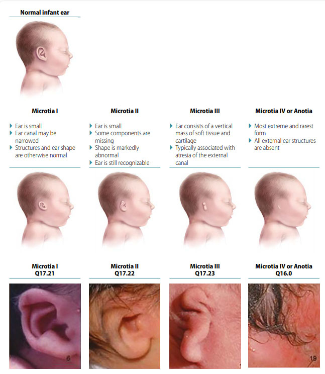
Microtia/anotia is a congenital malformation of the ear in which the external ear (auricle) is underdeveloped and either abnormally shaped (microtia) or absent (anotia). The external ear canal may be atretic (absent). The spectrum of severity in microtia ranges from a measurably small external ear (defined as longitudinal ear length below minus two SD from the mean, or approximately 3.3 cm in the term newborn) with minimal structural abnormality, to an ear that consists of few rudimentary structures and an absent or blind-ending external ear canal.
Fig. 13. Microtia/anotia

- Severity – I-IV degree, based on the extent of external ear involvement and atresia of the external canal.
- Sidedness – unilateral (usually unilateral; more often on right side) versus bilateral; severity can vary between the left and right sides.
Diagnosis
Prenatal. Microtia/anotia is easy to miss prenatally. Delineating the position and shape of the ear might require threedimensional ultrasound. Even if prenatal ultrasonography suggests microtia/anotia, the diagnosis should always be confirmed postnatally. When such confirmation is not possible – due, for example, to termination of pregnancy or unexamined fetal death – the programme should have criteria in place to determine whether to accept or not accept a case based only on prenatal data.
Postnatal. Microtia-anotia can be easily recognized and classified based on the newborn physical examination. However, abnormalities of the middle and inner ear, commonly associated with the more severe degrees of microtia, should be sought, and typically require advanced imaging (CT or MRI scan), surgery, or autopsy. Because microtia (second degree and above) is associated with hearing loss, hearing should be evaluated as soon as possible, ideally in the newborn period, so that appropriate management can be put in place.
Clinical and epidemiologic notes
- Hearing loss.
- Other anomalies and syndromes, especially those involving the mandible and face.
- Look for microtia/anotia occurring in conjunction with other anomalies and syndromes, especially those involving the mandible and face. Such conditions include the oculo-auriculo-vertebral spectrum (OAVS) and Goldenhar “syndrome” , as well as genetic syndromes, such as Treacher-Collins syndrome and trisomy 18, or teratogenic, such as retinoic acid embryopathy.
- Review examinations, procedures and imaging.
Checklist for high-quality reporting
| Microtia/Anotia – Documentation Checklist |
Describe in detail:
Take and report photographs: Show clearly the side and front; can be crucial for review. Describe evaluations to find or rule out related and associated anomalies:
Report whether autopsy (pathology) findings are available and if so, report the results. |