Congenital Anomalies of Genital and Urinary Organs: Hypospadias
Hypospadias is characterized by an abnormal (ventral) placement of the external urethral meatus in male infants. Normal placement of the urethral meatus is on the tip of the penis, whereas in hypospadias the meatus is ventrally and proximally displaced (on the underside of the penis).
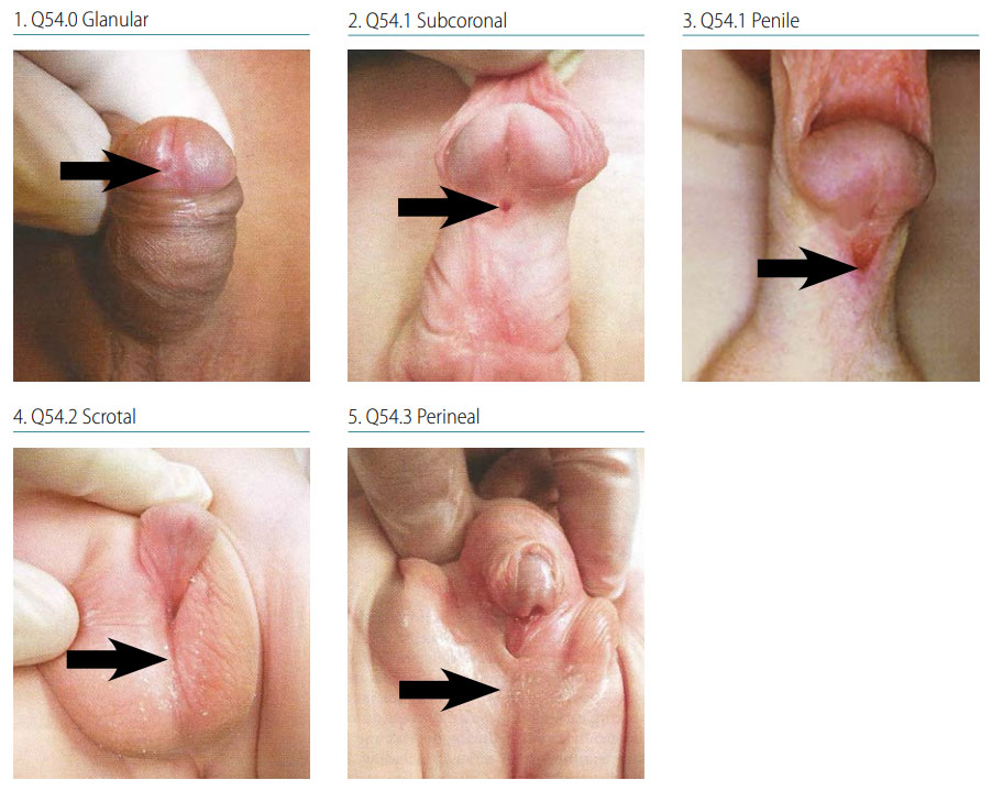
Fig. 32. Hypospadias

Photograph source: 1, 2, 4, 5: Nelson Textbook of Pediatrics (http://www.slideshare.net/wadoodaref/congenital-anomalies-videosession-v3-11680674).
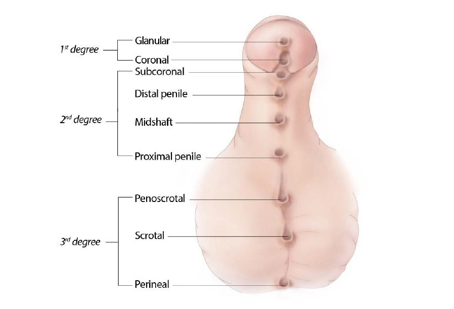
- Location: Hypospadias is classified by severity (see Fig. 32) depending on the location of the meatus – first degree includes the more distal forms, glanular and coronal; second degree includes subcoronal and penile shaft hypospadias; and third degree, the most severe, includes scrotal and perineal hypospadias.
- Potential associated findings: The shortening of the ventral side of the penis found in hypospadias can result in a curvature of the penis, known as chordee. Note the presence of chordee. Note if the testes are present or absent.
Diagnosis
Prenatal. Hypospadias is difficult to diagnose prenatally using ultrasound and may be confused with micropenis, penile cyst, chordee, or ambiguous genitalia. A prenatal diagnosis of hypospadias should always be confirmed postnatally.
Postnatal. A careful, systematic examination of the newborn should allow a firm diagnosis of hypospadias. Note that the milder forms such as glanular hypospadias are easily missed at delivery and may be discovered during circumcision. The surgical report may provide definitive detail of the urethral placement and whether chordee is present.
Clinical and epidemiologic notes
Hypospadias is often an isolated (>80%) non-syndromic anomaly.
- Note if testes are present/absent and if chordee is present.
- When stratified by type, the proportion of isolated defects decreases with increasing severity (penile 74% and scrotal 65%, the most severe).
- Look for additional anomalies, in particular for penile and scrotal hypospadias as these are associated with syndromes.
- Report all findings and obtain good clinical photographs (or note location of urethra in figure below) for the expert reviewer.
Checklist for high-quality reporting
| Hypospadias – Documentation Checklist |
Describe in detail:
Include photographs: Show clearly the location of the urethra or use drawing; can be crucial for review. Describe evaluations to find or rule out related and associated anomalies:
Report whether autopsy (pathology) findings are available and if so, report the results.  |
Note: Illustration indicates all possible locations for the malformation; select the location closest to what you see in the patient.